Use of Quadruple Zygoma Implants Technique for Atrophic Maxilla Rehabilitation with Immediate Loading - A Clinical Case Report
The maxilla with great atrophy can be due to various factors such as tumor resection, generalized aggressive periodontitis, genetic disorders or syndromes. The rehabilitation of patients with an atrophic edentulous maxilla presents a significant challenge for surgeons, and the treatment concept with zygomatic implants have evolved as an alternative for bone augmentation procedures. The aim of this review is to describe the rehabilitation of the maxillary with the use of four zygomatic implants (quad-zygoma implant technique). Within the limitations of this observation, we can state that full mouth rehabilitation with quadruple zygomatic implants technique can definitely be considered as a viable treatment option treating patients with atrophic maxilla. However, studies with more follow-up time and controlled clinical trials should be done in order to document the longevity of this kind of treatment modality.
Keywords: Atrophic Maxilla; Quad Zygomatic Implants; Immediate Loading
Atrophic maxilla can be due to various factors from bone loss, tumor resection or other genetic disorders and syndromes [1]. Fabrication of prosthesis with adequate retention and stability for patients with an atrophic edentulous maxilla presents a significant challenge. The aim of rehabilitation is not only to provide a cosmetically acceptable appearance, but also to restore oral functions, such as deglutition, mastication, and phonation [2]. Treatment options for edentulous patients include tissue-supported removable prosthesis, implant-supported removable prosthesis, and implant-supported fixed prosthesis. Implant-supported fixed prostheses have been proved to have high success rates in restoring facial esthetics, speech and function in these individuals [2,3]. The presence of inadequate bone quantity produce a potential problem for implant placement needing bone augmentation procedures. For decades, the atrophy of the maxillary process has been treated by large grafts from the iliac crest or calota in the form of onlays, in combination or not with a segmental osteotomy type Lefort; or by sinus inlays. Clinical studies with a long follow-up time reveal disparity in the survival of the implants after these procedures; Success depends to a large extent on the amount of preexisting bone in the residual bone crest and on the appearance of complications derived from the grafted area [4-10]. However, bone grafting procedures have few limitations like need for hospitalization, morbidity of donor site, unpredictable resorption of the bone graft, and delayed placement of the implant for the graft consolidation time [11-13].
Treatment concepts with zygomatic implants have evolved as an alternative for bone augmentation procedures. Combination of conventional implants and zygomatic implants has been used successfully for restoration of atrophic maxilla [5,14-20]. The placement of anterior conventional implants without grafting will be extremely difficult in severely atrophic premaxilla. In such situations, quadruple zygomatic implants can be used for the restoration of the edentulous maxilla [21-23]. This clinical case describes the restoration of the maxillary dentition using four zygomatic implants (quad zygomatic implant technique).
Female patient, 74 years old without major systemic disease, only slight hypertension but properly controlled. Presented an edentulous maxilla with resorption and with a complete prosthesis badly adapted with high mobility, reporting problems with poor chewing function and searching a treatment without the need of bone graft and the possibility to have a fixed restoration.
The same went to the Center of Dental Specialties to the service of Oral and Maxillofacial Implantology (Cibumax) for examination, diagnosis and resolution of problems.
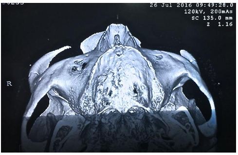
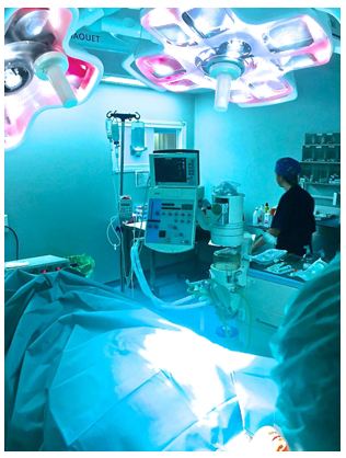
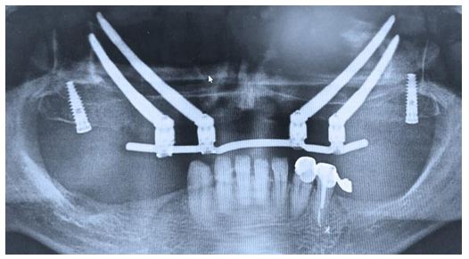
After the clinical, radiographic examination and definition of the treatment plan to be performed, a conscious sedation in hospital was improved with midazolam, was infiltrated 4% articaine and bupivacaine anesthetic for nerve block (Figure 1 and 2).
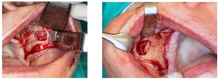
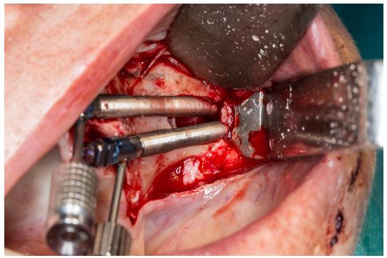
Crestal, anterior, and posterior vestibular releasing incisions were made, and mucoperiosteal flap was elevated to expose the alveolar crest, the lateral wall of the maxillary sinus, infra orbital nerve and the inferior rim of the zygomatic arch. A small window was made on the lateral wall of the sinus and detachment of the sinus membrane to assist visualization of drills during osteotomy preparation. Sequential osteotomy was done with 2.9 mm round bur, 2.9 mm cylindrical bur and 3.5 mm pilot drill (Figure 3 and Figure 4).
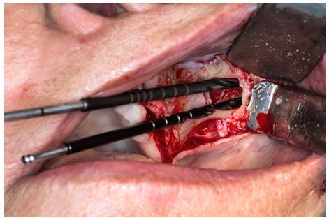
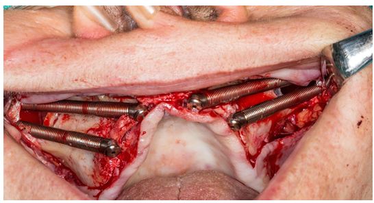
The anterior zygomatic implants Neodent® 4x52.5 mm were placed engaging the zygomatic bone with their platforms emerging at the canine region #13,23. The posterior zygomatic implants Neodent® 4x45 mm were placed through the infra-zygomatic crest emerging near the second premolar region #15,25; all zygomatic implants with a torque insertion of 65Nw (Figure 5 and 6). We placed 2 pterygoid implants Neodent® 4x17mm in #17, 27 position, with 30Nw of torque insertion for better support in the final restoration, we prefer not attached these pterygoids implants with the immediate loading for the low torque of them.
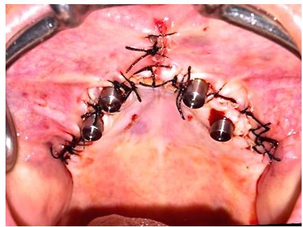
Straight multiunit abutments neodent® were placed for the preparation of a provisional complete fixed immediate load prosthesis, continuing with the closure flap sutures with black silk 000 (Figure 7).
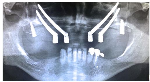
Impressions, bite registration tests and tooth color were taken for the preparation of the provisional immediate load prosthesis to be installed the same day of the implant surgery, and to do its respective postoperative radiographic record (Figure 8 and Figure 9).
The surgical procedure was performed without complications, achieving a good primary stability of the implants to be able to perform the provisional immediate load prosthesis and thus the patient reincorporate to their social life as soon as possible getting a comfort and aesthetics quite pleasant.
Zygomatic implants in the atrophic maxilla have been reported to be clinically successful [8-15]. Considering the high success rate of zygomatic implants and the need for immediate fixed prosthesis without grafting procedures, the use of four zygomatic implants for a maxillary fixed prosthesis can be a valuable treatment option [22-24].
The placement of double zygomatic implants bilaterally is well documented in the literature, with an extremely low implant loss rate [10,24-27].
This clinical report suggests that the placement of 4 zygomatic implants with immediate occlusal loading is a viable and effective treatment option for the rehabilitation of severely resorbed maxillae alternatively to maxillary reconstruction with autogenous bone grafts.
The anatomical configuration of the maxilla and zygomatic bone facilitated an extra sinus approach for placement of the zygomatic implants with a good primary stability, more than 60Nw of torque allowing the application of an immediate loading prosthesis.
The literature and our experience, shows that the technique with four zygomatic implants give good clinical results for the rehabilitation of atrophic jaws, with the added possibility of immediate loading of them and thus return the chewing and aesthetic function in a faster way to the use of bone grafts. This procedure requires through knowledge of the technique, superior surgical skills and meticulous training in prosthetic rehabilitation. We can state that full mouth rehabilitation with quadruple zygomatic implant technique can definitely be considered as a viable treatment option treating patients with atrophic maxilla. However, studies with more follow-up time and controlled clinical trials should be done in order to document the longevity of this treatment modality.